Hippocampus Atlas
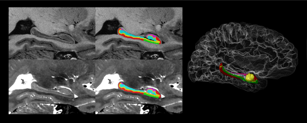
The hippocampus is a neuroanatomical structure that has been widely studied in the context of learning, memory, stress, and neurodegeneration. Neuroanatomically, the hippocampus is subdivided into several subfields with intricate morphologies and complex three-dimensional relationships.
To overcome the limited availability of post-mortem specimens and expertise in state-of-the-art high-field imaging, we conducted a coupling of MR acquisition and detailed segmentation techniques that allow for the reliable identification of hippocampal anatomy (including subfields). High-resolution and -contrast T1- and T2-weighted image volumes were acquired from 5 volunteers (2 male; 3 female; age range: 29–57, avg. 37) using a clinical research-grade 3T scanner and have final super-sampled isotropic voxel dimensions of 0.3 mm.
For more information and downloads, see the CoBrAlab website.
Cerebellum Atlas
We present a novel set of high-resolution in vivo atlases of the cerebellum developed by pairing MR imaging with a carefully validated manual segmentation protocol.
The cerebellum has classically been linked to motor learning and coordination. However, there is renewed interest in the role of the cerebellum in non-motor functions such as cognition and in the context of different neuropsychiatric disorders. The contribution of neuroimaging studies to advancing understanding of cerebellar structure and function has been limited, partly due to the cerebellum being understudied as a result of contrast and resolution limitations of standard structural magnetic resonance images (MRI). These limitations inhibit proper visualization of the highly compact and detailed cerebellar foliations.
For more information and downloads, see the CoBrAlab website.